Showing posts with label Hospitals of India. Show all posts

Friday, September 2, 2011

Enlarged heart (Cardiomegaly) : Causes, Symptoms, Tests & Diagnosis

Enlarged heart (cardiomegaly) isn’t a disease, but rather a symptom of another condition.
The term “cardiomegaly” most commonly refers to an enlarged heart seen on chest X-ray before other tests are performed to diagnose the specific condition causing your cardiomegaly. You may develop an enlarged heart temporarily because of a stress on your body, such as pregnancy, or because of a medical condition, such as the weakening of the heart muscle, coronary artery disease, heart valve problems or abnormal heart rhythms.
While having an enlarged heart may not always be preventable, it’s usually treatable. Treatment for enlarged heart is aimed at correcting the underlying cause. Treatment for an enlarged heart can include medications, medical procedures or surgery.

Symptoms:
In some people, an enlarged heart causes no signs or symptoms. Others may have these signs and symptoms:
  • Shortness of breath
  • Dizziness
  • Abnormal heart rhythm (arrhythmia)
  • Swelling (edema)
  • Cough
  • Chest pain
Causes:
Conditions associated with an enlarged heart include:
  • High blood pressure. Having high blood pressure can make it so that your heart has to pump harder to deliver blood to the rest of your body, enlarging and thickening the muscle.
  • Heart valve disease. Four valves within your heart keep blood flowing in the right direction. If the valves are damaged by such conditions as rheumatic fever, a heart defect, infections (infectious endocarditis), connective tissue disorders, certain medications or radiation treatments for cancer, your heart may enlarge.
  • Disease of the heart muscle (cardiomyopathy). Cardiomyopathy is the thickening and stiffening of heart muscle. In early stages of cardiomyopathy, you may have no symptoms. As the condition worsens, your heart may enlarge to try to pump more blood to your body.
  • Heart attack. Damage done during a heart attack may cause an enlarged heart.
  • A heart condition you’re born with (congenital heart defect). Many types of congenital heart defects may lead to an enlarged heart, as defects can affect blood flow through the heart, forcing it to pump harder.
  • Abnormal heartbeat (arrhythmia). If you have an arrhythmia, your heart may not pump blood as effectively as it would if your heart rhythm were normal. The extra work your heart has to do to pump blood to your body may cause it to enlarge.
  • High blood pressure in the artery connecting your heart and lungs (pulmonary hypertension). If you have pulmonary hypertension, your heart may need to pump harder to move blood between your lungs and your heart. As a result, the right side of your heart may enlarge.
  • Low red blood cell count (anemia). Anemia is a condition in which there aren’t enough healthy red blood cells to carry adequate oxygen to your tissues. Left untreated, chronic anemia can lead to a rapid or irregular heartbeat. Your heart must pump more blood to make up for the lack of oxygen in the blood when you’re anemic. Rarely, your heart can enlarge if you have anemia for a long time and you don’t seek treatment.
  • Thyroid disorders. Both an underactive thyroid gland (hypothyroidism) and an overactive thyroid gland (hyperthyroidism) can lead to heart problems, including an enlarged heart.
  • Excessive iron in the body (hemochromatosis). Hemochromatosis is a disorder in which your body doesn’t properly metabolize iron, causing it to build up in various organs, including your heart muscle. This can cause an enlarged left ventricle due to weakening of the heart muscle.
  • Rare diseases that can affect your heart, such as amyloidosis. Amyloidosis is a condition in which abnormal proteins circulate in the blood and may be deposited in the heart, interfering with your heart’s function. If amyloid builds up in your heart, it can cause it to enlarge.
Complications of enlarged heart can include:
  • Heart failure. One of most serious types of enlarged heart, an enlarged left ventricle, increases the risk of heart failure. Heart failure occurs when your heart can’t pump enough blood to meet your body’s needs. Over time, the heart can no longer keep up with the normal demands placed on it. The heart muscle will weaken, and the ventricles stretch (dilate) to the point that the heart can’t pump blood efficiently throughout your body.
  • Blood clots. Having an enlarged heart may make you more susceptible to forming small blood clots in the lining of your heart. If clots are pumped out of the heart and enter your circulatory system, they can block the blood flow to vital organs, including your heart and brain causing a heart attack or stroke. If clots develop on the right side of your heart, they may travel to your lungs, a dangerous condition called pulmonary embolism.
  • Heart murmur. For people who have an enlarged heart, two of the heart’s four valves — the mitral and tricuspid valves — may not close properly because they become dilated, leading to a backflow of blood. This flow creates sounds called heart murmurs. Heart murmurs are not necessarily harmful, but they should be monitored by your doctor.
  • Cardiac arrest and sudden death. Some forms of enlarged heart can lead to disruptions in your heart’s beating rhythm. Some of these heart rhythms are too slow to move your blood, and some are too fast to allow the heart to beat properly. In either case, these abnormal heart rhythms can result in fainting or, in some cases, cardiac arrest or sudden death.
Tests and Diagnosis :
If you have symptoms of a heart problem, your doctor will perform tests to determine if your heart is enlarged and to find out the underlying cause of your condition. In addition to a physical exam, these tests may include:
  • Chest X-ray. X-ray images help your doctor see the condition of your lungs and heart. If your heart is enlarged, it might first be detected by a chest X-ray, but other tests will usually be needed to find out the specific cause. Your doctor can also use an X-ray to diagnose conditions other than enlarged heart that may explain your signs and symptoms.
  • Electrocardiogram. This test records the electrical activity of your heart through electrodes attached to your skin. Impulses are recorded as waves and displayed on a monitor or printed on paper. This test helps your doctor diagnose heart rhythm problems and damage to your heart from a heart attack as well as give clues to other types of heart disease.
  • Echocardiogram. An important test for diagnosing and monitoring an enlarged heart is the echocardiogram. An echocardiogram uses sound waves to produce a video image of your heart. In this test, all of the chambers of the heart can be evaluated to determine if you do have an enlarged heart and to try and determine the cause. This test determines how efficiently your heart is pumping, assesses your heart valves, can look for evidence of previous heart attacks and can determine if you have congenital heart disease.
  • Stress test. A stress test, also called an exercise stress test, is used to gather information about how well your heart works during physical activity. Because exercise makes your heart pump harder and faster than it does during most daily activities, an exercise stress test can reveal problems within your heart that might not be noticeable otherwise. An exercise stress test usually involves walking on a treadmill or riding a stationary bike while your heart rhythm, blood pressure and breathing are monitored.
  • Cardiac computerized tomography (CT) or magnetic resonance imaging (MRI). In a cardiac CT scan, you lie on a table inside a doughnut-shaped machine called a gantry. An X-ray tube inside the machine rotates around your body and collects images of your heart and chest. In a cardiac MRI, you lie on a table inside a long tube-like machine that produces a magnetic field. The magnetic field aligns atomic particles in some of your cells. When radio waves are broadcast toward these aligned particles, they produce signals that vary according to the type of tissue they are. Images of your heart are created from these signals, which your doctor will look at to determine the cause of your enlarged heart.
  • Blood tests. Your doctor may order blood tests to check the levels of certain substances in your blood that may signal that you have a heart problem. Blood tests can also help your doctor rule out other conditions that may cause your symptoms.
  • Cardiac catheterization and biopsy. In this procedure, a thin tube (catheter) is inserted in your groin and threaded through your blood vessels to your heart, where a small sample (biopsy) of your heart can be extracted for analysis in the laboratory. Pressure within the chambers of your heart can be measured to see how forcefully blood pumps through your heart. Pictures of the arteries of the heart can be taken during the procedure (coronary angiogram) to ensure that you do not have any blockage.

Saturday, February 19, 2011

RapidArc IMRT - Advanced Radiation Technology for Cancer Treatment

Posted on February 20, 2011 by HealthnHeal


  • Comprehensive Cancer Care Under One Roof
  • Free Expert Medical Opinion from India's Top Cancer Specialists
  • Affordable, Low Cost Cancer Treatment Packages
  • Access to World Class Cancer Centers in India with best Doctors and nursing care
  • Highly advanced technology with Cyberknife, Linear Accelerator, Gama Knife etc.
  • Free guidance on Medical Travel to India
  • Free Airport Pick-up and Drop

RapidArc IMRT -The technology, described as revolutionary, was introduced for the first time ever in the U.S. in May 2008. It will change the way radiotherapy is planned and delivered. It is a major advancement from conventional Intensity Modulated Radiotherapy Technology in that it improves dose conformity while significantly shortening treatment times compared to conventional IMRT techniques. While ensuring all the benefits of IMRT, RapidArc delivers treatment within two minutes with precision, enabling physicians to improve the standard of care and treat more patients.


In a typical IMRT procedure, the gantry of the linear accelerator rotates to several ‘fixed’ beam angles. To treat the prostate, usually five to seven specific angled beams are used, each delivering a fraction of the total radiation for that day. Since these fields are limited to certain angles, the ability to avoid or minimise radiation to normal tissue is also limited. In the RapidArc procedure, there are no angles in which the beam stops. The treatment is delivered in one single rotation of the gantry around the body. There is no limitation to where and when the beam will be ‘on’ or ‘off,’ or how fast or slow it is being delivered. So, medical physicists and radiation oncologists will have nearly unlimited choices and degrees of freedom to design sophisticated and highly conformal treatment. This will result in less normal tissue damage in the high-dose treatment fields and fewer side-effects.

Previous approaches to arc IMRT therapy were restrictive, limited as they were by the design of the machine. RapidArc technology delivers precisely sculpted three-dimensional dose distribution with a single 360-degree rotation of the linear accelerator gantry. RapidArc uses Dynamic Multi Leaf Collimator, a variable dose rate, and a variable gantry speed to generate IMRT-quality dose distribution in a single optimised arc. Clinicians can thus deliver a continuously modulated dose to the entire tumour volume while sparing relatively normal or healthy tissue.

With RapidArc, a medical linear accelerator can target radiation beams at a tumour while making one continuous rotation around the patient. Conventional IMRT treatments are slower and more difficult for radiotherapy technologists because they target tumours using a complex sequence of fixed beams from multiple angles.

RapidArc differs from existing techniques such as helical IMRT or intensity-modulated arc therapy as it delivers a dose to the whole volume, rather than slice by slice. A single arc can deliver essentially similar dose distributions compared with IMRT plans. The treatment planning algorithm ensures treatment precision.

RapidArc radiotherapy technology advances the standard of care even in complex treatment plans. For example, in a case of multi-target cancer of the nasopharynx, RapidArc plans were found to be equivalent or better at target coverage, and superior in protecting critical structures including the spinal cord, the brain stem, the eyes, the optic nerve and chiasm, the parotid glands and the brain. RapidArc allows more control to tailor the dosage to suit the size, shape, and location of a tumour. Laboratory studies suggest that faster dose delivery may kill some cancer cells more effectively.

Faster treatment also contributes to precision by reducing the time for motion within anatomical structures. The technique reduces the probability of intrafractional tumour movements caused by the patient moving during treatment. Repositioning can make the treatment even longer.

RapidArc delivers volumetric IMRT in a single arc or multiple arcs of the equipment around the patient and makes it possible to deliver advanced image-guided IMRT two to eight times faster than is possible with conventional IMRT. Radiotherapy studies correlate the ability to spare more healthy tissue with fewer complications and better outcomes. RapidArc delivery produces as little as 400 Monitor Unit per fraction. Fixed-field IMRT treatments of similar quality can generate 1,000 to 2,000 MU.

RapidArc’s single gantry rotation, besides speeding up delivery, may result in less leakage and scattered radiation and a lower overall peripheral dose than in the case of conventional IMRT treatments.


Patient care being at the heart of everything at the Cancer Institute, the decision to invest in this equipment was a straightforward one. The use of this speedier and state-of-the-art technology will enable the institute to treat more patients with less stress and more comfort to them.


For more information on affordable and high quality cancer treatment in India please contact us anytime.
info@hospitalsofindia.net
info@healthnheal.com www.healthnheal.com

Thursday, January 27, 2011

The World's Top Five Medical Tourism Destinations!!


Medanta1

India has once again been named as one of the top Medical Travel destinations for 2010 by Tourism Review and the Hotel & Resort Insider! The country received well over a million medical tourists last year and that number is set to grow even further this year. With world class JCI, ISO and NABH accredited hospitals, the country is cementing itself as the preferred choice for medical tourists. With all the leading hotel chains being represented in India, medical tourists are spoilt for choice! India is also listed by Lonely Planet as a tourism destination to visit for 2010 with sights and sounds to cater for all types tourists.

Hospitals of India has been in forefront in promoting India as a healthcare destination and has served clients from well around the world! It is supporting efforts by the Indian Government under the Government Transformation Programme (GTP) to increase the number of medical tourists and to make India the number one hub for medical tourists!!

(Picture Above: Medanta – The Medicity, 1600 Bed Super Specialty Hospital)

See article below!

Medical Tourism & Travel: The World's Top Five Destinations |

Saturday, January 15, 2011

Why the aspirin is a wonder drug

Danny Buckland, Reposted On Saturday, January 15, 2010

It’s usually found in an unobtrusive white bottle in most bathroom cabinets. But the humble aspirin is being hailed as a medical miracle.

Breakthrough research this week showed aspirin could drastically reduce the risk of dying from many common cancers.

It’s staggering range – helping headaches through to critical conditions – means it could save millions of lives.

The British study proved that low doses of aspirin can cut death rates from cancers by about 30%, and up to 60% with prolonged use.

Professor Peter Rothwell’s team at the John Radcliffe Hospital, in Oxford, astonished the world with results from eight long-term studies involving 26,000 patients.

It reported that after five years, cancer death rates were cut by an average of 34% and up to 54% in gastrointestinal cancers. The results were seen across lung, pancreatic and oesophagus cancer as well as, to a lesser extent, brain cancer.

It is “the most amazing drug in the world”, according to Professor Alastair Watson, a consultant gastroenterologist at the University of East Anglia. “Penicillin has probably saved more lives but in terms of broad applications I don’t think anything comes close to aspirin. And it doesn’t cost much,” he said.

“Developed from willow bark, it started out as a painkiller which was good for fever and rheumatism. Then, about 20 years ago, it became clear that it was good for heart ­diseases and stroke.

“We then got to a position where virtually every patient who has had a heart attack, heart surgery, a stroke or problems with circulation in their legs is on aspirin. But to everyone’s astonishment and delight aspirin is going that extra mile.”

For pain relief, it penetrates the pain centres in the brain and switches them off while also reaching the nerve endings to neutralise the pain response mechanism. For cancers, it inhibits the damaging rogue cells by cutting off their blood supply. “It has been around for 100 years and was sold before the times of clinical trials and it is interesting to ask if it would be licensed today because of its side effects,” added Professor Watson.“Official government figures show it caused some 22 deaths in one year.

Many more had serious illness and most hospitals admit people with aspirin side effects almost every day. To put those figures into perspective, 70,000 people died from the effects of heroin in the same period.

“Aspirin is not the most dangerous drug but you cannot say it is completely safe.”

Experts caution against all middle-aged people – who stand to benefit most from the cancer application – immediately popping pills on a daily basis, and advise anyone with health concerns to get advice from their GP. But for a million Brits, the everyday ritual of swallowing a baby aspirin has life-saving implications.

“It’s about a quarter the dose you take for a headache,” said Mick Henderson, director of the Aspirin Foundation, the information service supported by Bayer, the company that developed aspirin.

He said: “Most GPs will put patients on to an aspirin if they have had a heart attack but more and more are putting them on to aspirin if they believe they are at risk from being overweight, smoking, stress or have a family history of heart attack at a relatively young age. So it can be taken even before heart attack."

“A lot of people are simply saying it is a wonder drug.”

Source: Daily Mirror Pic: Simon Stratford

Wednesday, December 22, 2010

US based Indian creates first artificial implantable kidney

TNN, Dec 4, 2010, Source: Times of India

NEW DELHI: US-based Indian origin researcher Shuvo Roy has created the world's first implantable artificial kidney. What's sensational about Roy's creation is that the organ, no larger than a coffee cup, will be able to mimic the kidney's most vital functions like filtering toxins out of the bloodstream, regulate blood pressure and produce the all- important vitamin D.
The artificial kidney has been tested successfully on a small number of animals. Large-scale trials on animals and humans are expected over the next five years. Once available, and if affordable, this creation by the Roy-led team at University of California will do away with the need for kidney dialysis.
This will be a boon for all patients with chronic kidney disease (CKD). At present in India, of the 150,000 new patients who suffer from end-stage renal failure annually, only 3,500 get kidney transplants and 6,000-10,000 undergo dialysis. The rest perish due to an acute shortage of dialysis centres and nephrologists to man them.
CKD is rising at a rapid pace in India and the majority of those who perish are either unable to find a suitable organ for transplantation or are unable to pay for the high dialysis costs.
According to Roy, the device has a filtration section to remove toxins from the blood, alongside a compartment with renal cells to conduct other functions of a kidney. He believes the artificial kidney could last for decades and require no pumps or batteries. Patients wouldn't require anti-rejection drugs (as is required after transplants) either because there would be no exposed natural tissues for the immune system to attack.
The University of California team is awaiting approval to conduct larger scale animal and human trials. Already, it has successfully tested the implant in a few rats and pigs.
"The payoff to the patient community is tremendous," said Roy. "It could have a transformative impact on their lives...With the right financial support, I think we could reach clinical trials in five years. But it's hard to say how long after that it becomes commercially available due to the uncertainties of the FDA and commercialization prospects.''


So what would this artificial kidney mean for developing world? ''It will be a real boon,'' said Dr S C Tiwari, director of nephrology and renal transplantation medicine at Fortis health care. He added: ''The biggest problem with CKD patients in India is that majority of them are diagnosed in the final stages where they would either require constant dialysis or a transplant. They would require dialysis three times week. However, of the 200,000 CKD patients requiring dialysis, only 10,000 get it, mainly because they can't afford it. Maybe only 1,000 such patients get it for free or at a subsidized rate in government hospitals. The artificial kidney, when available and if affordable, will be a miracle.'' Dr Madan Bahadur, nephrologist with Mumbai's Jaslok Hospital added, ''Work on creating tubular cells (that perform the biochemical work of the kidney) began a decade back. But bio-chemical engineering has so far not managed to replicate the kidney.''
According to Dr Jitendra Kumar, head of nephrology at Asian Institute of Medical Sciences, the main reason why this artificial kidney will be a real breakthrough is because it will be able to mimic the vital functions of a kidney like regulate BP and produce vitamin D — things a dialysis can't do.

Tuesday, October 26, 2010

Indian Hospital Accreditation Programme Goes Global

India’s national hospital accreditation programme- National Accreditation Board for Hospital (NABH), run and managed by the Quality Council of India, is going global. 55 hospitals of India have formally received NABH accreditation while another 365 have applied for it. Accreditation remains a voluntary process in India.
NABH will begin in the Philippines and plans to extend into Bangladesh and Nepal by the end of 2010, and into Sri Lanka in 2011. Future plans are to extend into Middle Eastern countries, starting off with the state of Dubai. NABH also sees African countries such as Kenya and Nigeria as logical future destinations, as Indian hospital chains see great potential in Africa. NABH has set up a separate division to look after its overseas operation, National Accreditation Board for Hospitals and Healthcare Providers (NABH) International.
The driver to service international hospitals is the expansion of large Indian hospital groups into subsidiaries or joint ventures in an increasing number of countries globally. Girdhar Gyani of NABH explains, "The vision to go global has been there for a while but the opportunity arose when hospital managements which already have NABH accreditation in India started demanding accreditation for their overseas hospitals as well. We are ready for competition from the American, Canadian and other international accreditation agencies which are already present in the targeted countries."
Hospitals have been complaining that international accreditors in a competitive market are too expensive and inflexible on costs. NABH say that a three-year accreditation from JCI in the Philippines costs close to $46,000, while a comparable NABH accreditation costs a little over half that amount. Although initially aimed at hospitals and clinics, NABH International will also offer accreditation to spas and wellness centres.
Through its exclusive Philippine representative, HealthCORE, NABH International has been launched to improve conditions, systems, processes, and skills of hospitals and professionals in the country. Dr. Sanjiv Malik of NABH says, "Accreditation is a practical solution to one of the main issues in medical tourism, which is quality and safety assurance. NABH International allows hospitals to build credibility and confidence, which generates recognition among foreign patients, thereby promoting medical tourism." The Department of Tourism (DoT) believes that accreditation is the key for medical tourism to truly flourish in the Philippines, as it will recognize the Filipino healthcare providers to be at par with world-class standards.
NABH International’s accreditation process entails onsite visits to assess if standards on access, care of patients, management of medication, patient rights, and infection control are met. Standards on continuous quality improvement, good governance, facility safety, human resources, and information management system are evaluated by highly qualified and well-trained assessors who are doctors, healthcare administrators, or nursing supervisors.

Sunday, September 19, 2010

IVF – Treating IVF Failure


One of the most frustrating problems in infertility today is IVF failure - also called implantation failure. This refers to infertile patients who have undergone many IVF cycles and produced beautiful embryos - but the embryos have consistently failed to implant for unexplained reasons. 13-SM42648
Many patients who fail an IVF cycle often give up. Not only are they disappointed, they are also upset about the fact that their doctor does not usually give them a clear explanation for why the IVF cycle failed - or what they can do differently the next time, to improve their chances of success. While some stop for financial reasons, others come to the conclusion that they are destined to never have a baby; and admit defeat prematurely.

Hospitals of India partner Clinics/Hospitals pregnancy rates in patients who have failed IVF cycles elsewhere is very high, because we can transfer more embryos in difficult patients (unlike fertility clinics in UK and Australia, where the number of embryos which can be transferred is limited by law). While transferring more embryos does increase the risk of high-order multiple pregnancies, this risk is negligible in difficult patients (for example, the older women or women with previous failed IVF cycles). In our fertility clinic, we customise the number of embryos we transfer for each patient we treat, rather than just blindly follow a guideline (which has been laid down for the general population, without considering each individual's specific problem).

The other common reason for a failed IVF cycle is a poor ovarian response, which means patients get few eggs and few embryos. For these patients, we offer the option of aggressive super ovulation, with high doses of HMG, in order to help them grow more eggs, so we have more embryos to transfer.

For patients with a poor ovarian response, we also offer the option of GIFT - gamete intra fallopian transfer, in which we transfer the eggs and sperm directly into the fallopian tubes by performing a laparoscopy. This has a better pregnancy rate than IVF, because we put the eggs and sperm back where they belong - in the fallopian tubes, rather than in our incubator. ICSI

Sometimes the reason for IVF failure is because the embryo transfer was technically difficult, because of cervical stenosis. This means that the transfer is often traumatic, and is associated with bleeding. For these patients, if their fallopian tubes are open, we prefer doing a fallopian tube transfer ( ZIFT zygote intra fallopian transfer) so that we can bypass the cervix and place the embryos directly in the fallopian tubes. This ensures a very high pregnancy rate.

Another group of patients who often do poorly in other IVF clinics are those who have PCOD. Because many doctors are so worried about the danger of OHSS (ovarian hyper stimulation) in these patients, they often end up super ovulating these patients badly, and retrieve few poor quality eggs, compromising the pregnancy rate. In our fertility clinic, we prevent OHSS by carefully aspirating each and every follicle at the time of egg retrieval , and flushing it repeatedly with a double-lumen needle, until it collapses completely. By removing the follicular cells which are responsible for producing VEGF and causing OHSS, we have been able to prevent OHSS in PCOD patients very successfully in our fertility clinics/hospitals by using this novel technique.

Successful embryo implantation depends upon the health of the embryo, and one of the reasons embryos may fail to implant is that they may be chromosomally abnormal (even though they look normal). Research has shown that the incidence of chromosomal abnormalities even in good looking embryos is as high as 50% !

We can also offer the following advanced technique to help patients who have failed multiple cycles of IVF.

After In Vitro fertilization, which is performed in the normal fashion, we perform an embryo biopsy on Day 3, using a laser, and study the genetic composition of each embryo. This allows to select only the chromosomally normal embryos. The normal embryos are then transferred into the uterus on Day 5, when they are blastocysts.

This combined technique offers many advantages, especially for older women, who are more likely to produce abnormal embryos.

1. It allows us to select the chromosomally normal embryos. Not only does this increase the chances of embryo implantation, it also means the risk of a genetically abnormal baby is reduced.
2. We drill the zona with a laser. This allows us to facilitate embryo hatching , thus increasing the chances of embryo implantation.
3. Since we are transferring blastcysts on Day 5, the synchronisation between embryo and the endomterium is increased, thus enhancing implantation.
4. Since we can transfer fewer embryos ( each embryo now has a higher chance of becoming a baby ), the risk of multiple pregnancies is also reduced.
Fertility Mother Baby

Since this technique is very labour-intensive and technologically demanding, the cost is more than that of a regular IVF cycle. However, for patients who have failed 2 IVF cycles, and are not happy about the idea of repeating another similar IVF cycle again; and for older patients, this advanced option can be very cost-effective.

Thursday, September 16, 2010

Artificial Corneas Restore Sight for the First Time

Lab grown artificial corneas have been transplanted into patients' eyes for the first time, offering hope to millions of partially sighted people.
The new technique involves growing human tissue or collagen in the lab and then shaping it, using a contact lens mould.
Damaged and scarred tissue from the front of the eye is then removed and the "biosynthetic" replacement is stitched in its place, reports the Telegraph.
Ultimately, existing cells and nerves in the eye grow over the artificial cornea, incorporating it fully into the eye.
Globally, diseases that lead to clouding of the cornea affect more than 10 million people, making them the most common cause of blindness.
The first trials of the operation have shown that it is just as successful as live tissue transplantation and in some cases patients have had their sight fully restored.
May Griffiths, of Linköping University, Sweden, who led the study, said: "We were very excited by the results. The study is the first to show that an artificially fabricated cornea can integrate with the human eye and stimulate regeneration."
"With further research, this approach could help restore sight to millions of people who are waiting for a donated human cornea for transplantation," Griffiths added.
The cornea is a vulnerable shield or lens protecting the eye and plays a key role in creating vision.
Patients did not experience any rejection reaction or require long-term immune suppression, which are serious side-effects associated with the use of human donor tissue.
The bio-synthetic corneas also became sensitive to touch and began producing normal tears to keep the eye oxygenated.


Friday, August 27, 2010

Procedures at Hospitals of India

Dental

Dental

Dental care is an excellent option for the medical tourist in most cases, although certain procedures like braces may be inappropriate based on the repeated visits necessary for adjustments. In most cases though, appointments and lab work can be expedited to meet your time-line, and treatments can easily be combined with sightseeing and a holiday for maximum benefit.

IVF - Fertility

IVF – Fertility

Infertility is defined as the inability to get pregnant after one year of trying and can be caused by many factors, both male and female. It is estimated that one in six couples are affected by infertility, and this number is increasing as some families delay childbirth. Fertility treatment, or infertility treatment, falls into one of three categories: ovulation induction, artificial insemination, or assisted reproduction.

Cosmetic Surgery

Cosmetic Surgery

Cosmetic surgery, also known as plastic surgery or aesthetic surgery, as the name suggests, does not always involve surgical procedures as in the case of Botox injections or lip augmentation. Either way, the aim of cosmetic procedures is to enhance ones appearance towards a personal aesthetic ideal. The number of cosmetic surgeries in North America has increased over 50% in the last few years alone.

Surgery3-150x150

CCSVI (Liberation Treatment)

At Hospitals of India we are always looking to add procedures in our many destinations to give our clients the choices they desire. We are pleased and excited to offer MS – CCSVI Treatment to our ever growing list of procedures.


Orthopaedics

Orthopaedics

Orthopaedics is the branch of medicine dealing with the skeletal system. The work of an orthopaedic surgeon may range from hand reconstruction to full joint replacements such as knees and hips. The orthopaedic surgeon is also skilled at using foreign materials such as pins and screws to hold bones to their proper alignment.


Bariatrics (Weight Loss)

Bariatrics (Weight Loss)

Bariatric medicine is the study and treatment of obesity and the health conditions associated with it. Many health conditions are associated with obesity including type II diabetes, high blood pressure, gout, and heart and breathing disorders, to name a few.


Ophthalmology

Ophthalmology (Eye Surgery)

Ophthalmologists specialize in the medical and surgical treatment of eye disorders, vision measurements for glasses (refraction), eye muscle exercises (orthoptics), and the prevention of blindness and care of the blind.


Other ProceduresOther Procedures

At Hospitals of India we are always looking to add procedures in our many destinations to give our clients the choices they desire. If there is a procedure that you are interested in and you don’t see it here please ask us. There are so many different procedures available it is difficult to have all of them listed. Our network of Hospitals offer pretty much any procedure available. So please let us know how we may help you get the information you desire.

Saturday, August 21, 2010

Hip Resurfacing in India

Birmingham Hip Resurfacing in India 

Categories: Health, News, OrthopedicsBirmingham_Hip_Resurfacing
Tags: Health Care, Medical, Orthopedic Surgery

Is hip resurfacing right for me?  

Hip resurfacing is a surgical procedure designed to conserve more bone than the traditional total hip replacement (THR). It allows most people to return to the activities they once enjoyed prior to experiencing hip problems.

The Birmingham Hip Resurfacing (BHR) System, registered trademark of Smith & Nephew, is the first FDA-approved hip resurfacing system in the U.S., and since its introduction in 1997 the procedure has helped to cure an estimated 60,000 patients worldwide.

“Hip resurfacing allows most people to return to any activities they enjoyed prior to their hip problems.”

What is Birmingham Hip Resurfacing?

bhr_diagram

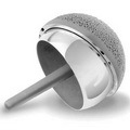
The Birmingham Hip is a two-part system that uses durable high-carbide cobalt chrome to resurface the ball and socket joint.

Unlike the THP – which requires the upper portion of the femur bone be cut off in order to accept the stem of the THP device – the BHR resurfaces the femoral ball with a metal cup. The pelvic socket or acetabulum is also resurfaced to create a metal-on-metal joint.

Smoothing the worn areas of the ball-and-socket joint with the high-carbide cobalt chrome eliminates pain and lets the hip move naturally.

Advantages

The main advantage of hip resurfacing is that the procedure conserves the complete femur bone, preserving the patient’s future option to undergo a THP if needed.

  • Less bone removal
  • Decreased risk of hip dislocation
  • More natural movement
  • Greater range of motion

Disadvantages

The potential disadvantages of hip resurfacing include:

  • Femoral neck fractures (rate of 0-4%)
  • Aseptic loosening
  • Metal wear
  • Metal ions in the blood and urine

Best Candidates for Hip Resurfacing

Simply put, hip resurfacing is intended for younger patients who are not overweight. Ideal candidates including those who:

  • Suffer from osteoarthritis
  • Have strong healthy bones
  • Are Under 60
  • Do not have an infection
  • Are not allergic to the metals used

The surgery is typically more suited to male patients, however females who best match the ideal requirements may be considered.

While hip resurfacing is a great alternative to THP surgery, it’s simply not suitable for everyone. Those who may not be suitable for hip resurfacing include patients who suffer from:

  • Severe bone loss in their femoral head
  • Large femoral neck cysts
  • Femoral head cysts larger than 1cm
  • Rheumatoid arthritis
  • Osteonecrosis (poor blood supply to the femoral head)
  • Obesity
  • Diabetes
  • Impaired kidney function

People who are tall, thin or small boned will also be advised not to undergo the procedure.

To ensure the long-term success of your treatment it’s essential to chose an experienced surgeon and take their advice.

Recovery

In most cases patients return home after 1-4 days. You may begin putting weight on the leg immediately however your doctor may advise using a cane or crutches for the first few days or weeks.

A physiotherapist will give you exercises to help increase your range of motion, and you’ll be required to visit the orthopedic surgeon for regular check-ups.

Most patients resume regular activities after around 6 weeks.

hip-resurfacing-x-ray

X-Ray of Brimingham Hip Resurfacing Surgey

Results

A typical hip resurfacing will last a long time. Studies have shown a 98.4% revision-free survival of the surgery at the five year mark, and a 95% revision-free survival at 10 years – comparable to that of a traditional total hip replacement in the under-60 age group.

Since hip resurfacing was introduced in 1997, many athletes have opted for the procedure and gone on to compete at an international level. They include Ron Noreman 2010 winner of NPC Empire States Bodybuilding Championships and 2006 Tour de France winner Ian MacLaren.

If you are considering Birmingham hip Resurfacing or any orthopedic surgery, visit Hospitals of India to discover an affordable alternative that does not require you to have insurance while also enjoying the benefits of traveling overseas to India. Visit top orthopedic centers at Hospitals of India and have consultations with top board certified orthopedic surgeons, led by famous orthopedic surgeon Dr. Ashok Rajgopal, Chairman, Medanta Institute of Bone & Joint. 

Wednesday, August 18, 2010

Range of Surgical Options for Knee Surgery


Total Knee Replacement

Total Knee Replacement (TKR), also referred to as Total Knee Arthroplasty (TKA), is a surgical procedure where worn, diseased, or damaged surfaces of a knee joint are removed and replaced with artificial surfaces. It is a common surgical procedure most often performed to relieve the pain and disability from degenerative arthritis, meniscus tears, osteoarthritis, cartilage defects, and ligament tears. The most common cause for knee pain which needs Knee Replacement Surgery is osteoarthritis. In Total Knee Replacement, the surgeon removes damaged cartilage and bone from the surface of your knee joint and replaces them with a prosthesis/implant of metal and plastic. Total knee replacement can help put an end to arthritic pain in your knee and enable you to resume a functional and active lifestyle.


Partial Knee Replacement


Most people are aware of the total knee replacement surgery. This involves replacing the unhealthy surface of the entire knee joint with metal and plastic implants. It is a very successful operation with good long term results. However a large percentage of patients have arthritis limited to one part of the joint alone. Replacing the whole joint in these patients is overkill and unnecessary. Many middle aged men and women develop osteoarthritis of the knee. Osteoarthritis of the knee affects the inner half or medial compartment to start with and then proceeds to affect the outer half or lateral compartment. In this operation only that part of the knee, which is unhealthy, is replaced. The normal surfaces are left alone. This operation has several advantages over total knee replacement surgery like small incision, longer life of implant and post operative hospitalization is reduced and return to normal is much faster than total knee replacement surgery.


Simultaneous Bilateral Kn
ee Replacement

A simultaneous procedure means that both knees are replaced at the same surgery, in one day. The benefit of simultaneous knee replacement is that both problems are taken care of at one time. The overall stay and rehabilitation can be done in a shorter time, and there is only one hospitalization. Patients also only require one anaesthesia.


Revision Knee Replacement


Total Knee Replacements that need to be revised after 15-20 years (post primary surgery) are also done here. The most advanced prosthesis and Computer Assisted Surgery (CAS) is also used for Revision Knee Replacements.


Advanced Technology and Latest Implants


Computer Assisted Knee Replacement Surgery

In computer assisted Knee Replacement, the computer guides the surgeon in precisely planning the exact amount and angle of bone to be removed during surgery. This, in conventional method is done by simple eye-balling. This technology helps to perform the surgery through small incision (Minimally Invasive Surgery) with 100% precision, eliminating "Human Error" during surgery and helps in perfectly aligning and balancing of knee joint (with accuracy of 0.5 mm and 0.5 degrees). It also helps in increasing implant longevity (20-30 years).


Gender Specific Knee Surgery


A women's anatomy is clearly different than a man's anatomy. Research shows women and men are different all the way down to their knees. Taking this fact into consideration knee replacement implants have been designed based on an average size of women's and men's knees. Nowadays gender specific implants are extensively used in Knee Replacement Surgery for better results. . A gender-specific Knee implant is a prosthesis that is a great boon for women and specifically designed for their needs. High Flex Rotating knee Platform
The design of the high flex implants closely replicate normal knee. Rotating knee replacement implants can swing backward and forward giving the knee flexibility to move forward and outward. High flexion gives patient the freedom to the extent that the patient can even squat , cross leg and sit on the floor.


Physiotherapy and Rehabilitation


Physiotherapy and Rehabilitation services help the patient to return back to normal activity quickly after the surgery. The network hospitals have highly advanced Physiotherapy Unit and well experienced physiotherapists. Some of the sessions of physiotherapy is included in the package and if the patient wants extra sessions of physiotherapy then it is provided at a nominal cost. In your physical therapy sessions you will walk, using crutches or a walker, bearing as much weight as indicated by your doctor or physical therapist. You will also work on an exercise program designed to strengthen your leg and increase the motion of your knee.

Wednesday, August 4, 2010

Dr Ashok Rajgopal, MS, Mch( Liverpool)

The first Orthopaedic Surgeon in India to perform a bilateral Total Knee Replacement Surgery in 1987.
Appointed Honorary Orthopaedic surgeon to the President of India in 1997.
The first Orthopaedic Surgeon in India to perform a Unicompartmental Minimally Invasive Replacement in 2002.
Designer of the instrumentation used in Minmally Invasive Surgery for Zimmer.
Orthpaedic surgeon to several internationally acclaimed sportsmen like P.Gopichand, Baichung Bhutia, N.Kunjarani Devi etc. all of whom have gone on to win laurels for our country after undergoing surgery for sports related knee and ankle injuries.

Tuesday, August 3, 2010

New Found treatment for Multiple Sclerosis

Vein Angioplasty for Multiple Sclerosis:

The Italian researchers discovered that, in persons with multiple sclerosis, the veins which acted as the main drainage pathways for blood flowing from the brain back to the heart were substantially narrowed and even blocked. These included the jugular veins, veins along the spinal column, and other veins such as the azygous vein.

The persons with MS have impaired venous drainage from the brain and that such a problem caused the phenomenon of “reflux”. This means the venous blood would flow back toward the brain as it established new pathways around the blocked and narrowed veins creating symptoms for Multiple Sclerosis.

The suggested treatment basis on this discovery is a treatment is called "the liberation procedure”. The problematic veins are first identified by venography. Then, balloon angioplasty is used to open up the problematic veins and, in some cases, stents are inserted in non- responding sections. The procedure is relatively non-invasive and is done in day hospital under local anesthesia. Access to the veins is through the left femoral vein in the thigh. Total time in the hospital is usually less than 6 hours and the subject has a compression dressing on for 24 hours.

Procedure Cost - Just USD 5200!!

Kindly feel free to get in touch to know more about this procedure or to know more about Medical Facilities in India at info@hospitalsofindia.net